CorrSlide™


Correlative Microscopy Coverslip, ITO coated, with high precision fiducials


Correlative microscopy is a sophisticated approach combining the imaging capabilities of different, powerful but typically separated microscopy platforms. Laborious, time consuming and limited precision procedures of getting a sample from one instrument to another have been so far one of the biggest struggles in obtaining fast and reliable results. CorrSlide™ technology uses integrated, high precision fiducials which enable easy relocation of samples and finding of exactly the same spot on a sample with micron precision under different resolutions and imaging techniques. Transparent conductive oxide coating (ITO) allows sample investigation under light microscopy and supports charge drain in electron scanning microscopy. It is also compatible with cell cultures and suitable for cells growth. CorrSlide™ can be provided with or without fiducial marks.

Correlative Microscopy Coverslip, ITO coated, with high precision fiducials


Benefits

  • Transparent for light / fluorescence microscopy
  • Conductive for scanning electron microscopy
  • Fiducial marks for precise, manual or automated localization of region of interest
  • Faster retrieval of region of interest
  • Secures sample position in relation to fiducials
  • Enables finding of rare or targeted structures
  • Enables a retrieval of region of interest even without characteristic points on an image
  • Enables processing of sample between image acquisitions
  • Enables integration of imaging platforms
  • Facilitates merger of functional and structural information
  • Eliminates need of conductive over-coating for ultrathin samples


Application example: Overlay of light (red) and electron microscopy images allows for classification of neural processes and synapses.

Application example: Overlay of light
Image courtesy of R. Hahnloser, Institute of Neuroinformatics, University of Zurich and ETH Zurich.


Applications

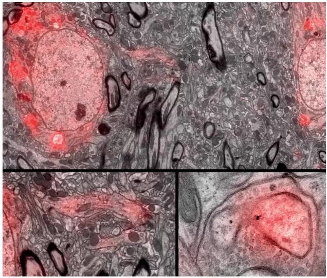
  • Light/fluorescence microscopy
  • Scanning electron microscopy (SEM)
  • Structured illumination microscopy (SIM)
  • Bioscience research
  • Imaging of cells, tissues, soft materials and proteins
  • Cell biology
  • Neurobiology
  • Studies of host-parasite interaction
  • Analysis of symbiotic relations


Technical Data

Substrate material

D263M glass (colorless borosilicate glass with very low iron content, meets requirements laid down in ISO 8255-1, has high spectral transmission, excellent flatness and a refractive index finely adapted to microscopes, very good resistance to chemical attack, fire polished)
Other materials on request


Substrate dimensions

22mm x 22mm x 0.17 ±0.005mm,

other dimensions and shapes on request


Fiducial marks (customer specific)

High precision
High contrast for excellent visibility
Flexible design adaptable to customer specific application


ITO coating

Premium quality, low defect, full-surface coating
High transparency in VIS range (Tavg>83% λ = 450-780 nm)
Optimized smooth surface texture
Compatible with cell cultures and suitable for cells growth


Other

Durable, unique serial number
Ultraclean surface
Packed under clean-room (class 100) conditions